marketing@innermed.com


Endoscopy Clinical Case: Brucella's gland hyperplasia and cyst
▶ Female, 54 years old;
▶ Findings under Endoscopy:
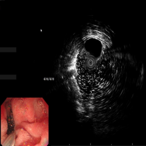
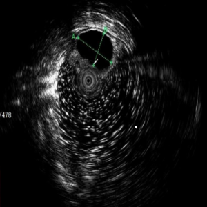
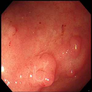
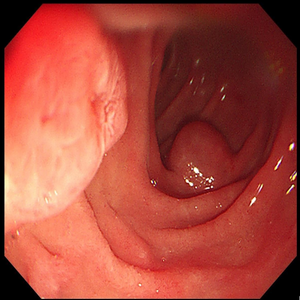
The esophageal mucosa is smooth, the vascular network is clear, and the cardia is normal. Gastric mucus is clear with medium amount. The gastric fundus and gastric body had multiple round-shaped white bulges with a diameter of about 0.2 cm, and scattered polypoid lesions with a diameter of about 0.2-0.3 cm. The gastric body was scattered with old blood stains, and the folds were arranged regularly. The gastric angle is arc-shaped and symmetrical, and the mucosa is smooth. The gastric antrum mucosa was not smooth, red and white, mainly red, and an irregular lesion with light yellow color was seen on the anterior wall near the pylorus, and the mucosa was light yellow. The pylorus is round and well opened and closed. The duodenal lumen has no deformation, scattered irregular bulges in the mucosa, no abnormalities in the duodenal papilla; scanning with a mini probe EUS, a non-echo hemispherical bulge with a size of about 8.6*7.1cm in the descending duodenum segment can be detected, and the interior is transparent, and cystic fluid seems to be seen.


▶ Diagnosis:
Chronic non-atrophic gastritis;
Gastric polyps;
Multiple bulges in the stomach: Changes in Haruma Kawaguchi disease;
Duodenal Bulb Lesions: Brucella's gland hyperplasia;
Prominence of descending duodenum: cyst;
Mini-Probe EUS Clinical Applications
2023-06-20
Endoscopy Clinical Case: Esophageal bulge considered tumor
2023-06-20
Endoscopy Clinical Case: Rectal polyps (partial Ca change possible)
2023-06-20
Endoscopy Clinical Case: Brucella's gland hyperplasia and cyst
2023-06-20
Endoscopy Clinical Case: Colon cancer
2023-06-20