marketing@innermed.com


Endoscopy Clinical Case: Colon cancer
▶ Male, 48 years old
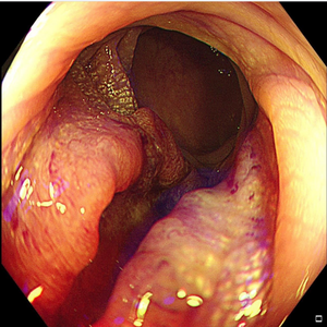
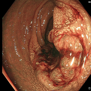
▶ Findings under endoscopy:
No obvious abnormalities were found in the terminal ileum. The opening of the appendix was half-moon-shaped and the lip of the ileocecal valve was smooth and smooth. The cecum, ascending colon, transverse colon, and descending colon have unobstructed intestinal lumen, normal shape of the intestinal valve, smooth mucosa, and an irregular new growth was seen at the junction of the straight and second colons about 18-20 cm away from the anus, with a depression at the top and obvious dilation of the glandular duct opening, deformation, partial fusion, and see thick twisted blood vessels. Scattered white flat polyps with a diameter of 0.2-0.3cm in the rectum were treated with APC cauterization and electrocoagulation respectively, and internal hemorrhoids were seen at the anus;


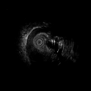
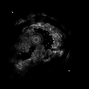
▶ Findings under mini probe EUS:
The lesion presents a hyperechoic mass shadow, mixed with hypoechoic shadows, originating from the mucosal layer, and invading the submucosa, muscularis mucosa, and full-thickness muscle propria.
▶ Diagnosis: colon cancer, multiple colonic polyps APC
Mini-Probe EUS Clinical Applications
2023-06-20
Endoscopy Clinical Case: Esophageal bulge considered tumor
2023-06-20
Endoscopy Clinical Case: Rectal polyps (partial Ca change possible)
2023-06-20
Endoscopy Clinical Case: Brucella's gland hyperplasia and cyst
2023-06-20
Endoscopy Clinical Case: Colon cancer
2023-06-20